Incontinentia pigmenti treatment is symptomatic and supportive. There is currently no cure for IP.
For infants and children, parents should engage a team of physicians to address the numerous features of the spectrum of IP. Preferably, these approaches should be guided by and supported by a geneticist familiar with the condition and its manifestations. The team may include a pediatric ophthalmologist and/or a pediatric retinal specialist, a neurologist, dermatologist, and eventually a pediatric dentist. Each specialist should determine what is in the child’s best interest for incontinentia pigmenti treatment depending on appropriate testing, including sedations of any kind.
One summary may be found here: National Protocol for the Diagnosis and Care of Rare Diseases: Incontinentia Pigmenti. For additional recommendations, jump to:
Eyes
Recommended Specialists – Pediatric Ophthalmologist and/or Retinal Disease Specialist, Ophthalmologist
Our IP experts created a list of incontinentia pigmenti care providers who specialize in treating eye issues.
The following treatment guidelines are a result of the NFED’s International Conference on Incontinentia Pigmenti (ICIP) in 2025. You can read a conference summary which was published in the American Journal of Medical Genetics. For the full eye treatment protocol, read Consensus Guidance for Screening, Classification, and Treatment of Retinopathy Associated with Incontinentia Pigmenti which was published in Ophthalmology Science.
Required Eye Examinations
It is imperative to have a complete medical eye examination as soon as possible after diagnosis.
A dilated retinal examination as soon after birth as the neonatologist or anesthesiologist thinks is safe. Sometimes, if there are any suspected retinal blood vessel abnormalities, an examination under anesthesia is required. The important thing is for the ophthalmologist to look at the optic nerve, the macula (in the center of the retina), and the far peripheral retina, where the typical pathologic events tend to occur. If retinal abnormalities are present, wide-field fluorescein angiography (WFFA) and treatment should follow as soon as possible, ideally within one month. If the ophthalmoscopic exam is normal, a screening WFFA should still be performed within three months, because many infants will have evidence of disease on WFFA that is not visible clinically.
A fluorescein angiogram is highly recommended, regardless of age, initially and at follow-up unless the retinal specialist decides the view of the retina is easily and completely obtainable without the angiogram. An angiogram will help the physician see the vessels located in the periphery of the eye where bleeding can occur and which cannot be seen by the naked eye.
The retinal specialist will determine how frequently to repeat the examinations and the angiograms depending on retinal findings obtained with routine examination techniques. As our technology evolves and the camera systems change, newer approaches to imaging may replace fluorescein angiography, including ocular coherence tomographic angiography (OCT-A).
These examinations and imaging systems are looking for serious abnormalities in the growth and development of the small blood vessels that nourish the retina, similar to those that occur in very low birth weight premature babies, called “retinopathy of prematurity”. Discovery of these changes requires monitoring for the further development of abnormal blood vessels, new blood vessel growths, and bleeding that results from them, which would then require treatment with laser or freezing therapies to obliterate the abnormalities to salvage the retina from further damage.
Severe retinal blood vessel disease is sometimes associated with blood vessel damage in the brain and thus is a marker to pursue imaging studies of the head. With respect to the eyes themselves, some babies with IP, and even some older patients, might benefit from laser treatment in an effort to prevent retinal detachment or vitreous hemorrhage from the consequences of the typical retinal neovascularization that occurs in this disorder.
Staging of IP-associated Retinopathy
The ICIP proposed the following consensus grading scheme for IP-associated retinopathy based on dilated ophthalmoscopic examination and fluorescein angiography (FA) (below table):
- Stage 0 (no retinal disease)
- Stage 1 (avascular retina or retinal vascular abnormalities, including arterial occlusion, with or without leakage)
- Stage 2 (extraretinal neovascularization)
- Stage 3 (retinal detachment, with or without foveal involvement)
Treatment and Monitoring Guidelines for IP-associated retinopathy
At the present time, there is no level 1, 2, or 3 evidence regarding the safety and efficacy of these therapeutic interventions, and level 4 evidence is also limited. The following treatment recommendations reflect the consensus of the ICIP participants developed to provide general guidelines for patients, families, and pediatricians for acceptable current practice based on available knowledge. The ICIP participants acknowledge that given the low prevalence of disease, it may never be possible to develop higher quality evidence, and that these practice patterns may evolve over time with new information.
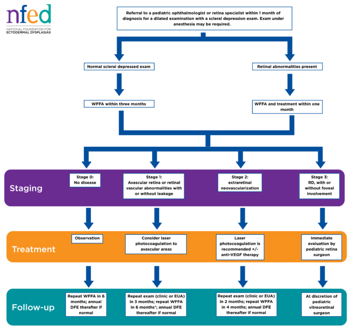
- Stage 0 (no retinal disease): Observation is recommended. To the experts’ knowledge, there have been no cases of disease progression following a normal wide-field fluorescein angiography (WFFA) at baseline. Nonetheless, given the rarity of the condition and the fact that image quality can be challenging especially in small eyes with low doses of fluorescein, it may be prudent to consider repeating WFFA within six months if there is any concern.
- Stage 1 (avascular retina or retinal vascular abnormalities with or without leakage): There are insufficient data to support the effectiveness of either anti-VEGF therapy or laser treatment as disease modifying interventions. However, many of the ICIP authors consider laser photocoagulation to avascular areas in these eyes given the long-term risk of avascular retina including the development of neovascular complications, especially if there is a risk of a patient being lost to follow-up. A subsequent follow-up examination should occur within three months (clinic or experimental autoimmune uveitis [EAU] at physician discretion) and repeat WFFA within six months.
- Stage 2 (extraretinal neovascularization): In the presence of neovascularization, most ICIP authors would recommend laser photocoagulation to areas of avascular retina, with some also recommending anti-VEGF therapy. Although spontaneous regression of neovascularization can occur in IP, laser may reduce the risk of both early progression to tractional retinal detachment (TRD). It is a serious condition where the retina is physically pulled away from the back of the eye by contracting scar tissue or abnormal blood vessels and later progression to rhegmatogenous retinal detachment (RRD). It is the most common type of retinal detachment, occurring when a tear or hole in the retina allows fluid to seep underneath, lifting the retina away from the supportive tissue and blood vessels behind it RRD. Closer follow-up is recommended given more advanced disease, with repeat exam within two months (clinic or EAU at physician discretion) and WFFA within four months. EAU is a widely used, induced T-cell-mediated autoimmune disease where a subject’s immune system attacks its own retinal and uveal tissues.
- Stage 3 (retinal detachment, with or without foveal involvement): Pediatric vitreoretinal surgical consultation and intervention is indicated as soon as possible.
Proposed screening and treatment protocol is detailed in the chart below.
If an important issue or question should arise at any time during an examination, immediately see a retinal specialist or pediatric ophthalmologist and refer back to the full ophthalmologic examination thereafter.
If any eye or head trauma occurs at any time throughout life, get an ophthalmic examination as soon as possible to rule out partial or full retinal detachment.
Additional Guidelines
Most people affected by IP have normal vision (with corrective lenses). Some problems, like near- and far- sightedness, are common in persons with IP. But, these are probably no more frequent than the general population without IP.
The classical eye finding in IP is an abnormality in the growth and development of blood vessels in the inside of the eye (the retina). Growth of abnormal blood vessels and the associated scarring can cause loss of vision, but can be treated if recognized early enough.
For this reason, babies diagnosed with IP should have the full IP eye examination immediately after birth and be followed by an ophthalmologist closely. Have a pediatric ophthalmologist or retinal disease specialist conduct a careful examination
In some older literature, some eye abnormalities have been reported, such as small eye (microphthalmos), cataract, degeneration of the optic nerve (optic atrophy) and even or total blindness. However, these complications occurred in an era before the retinal blood vessel abnormalities were recognized and were treated in the more recent times. However, a cataract is never a primary feature of IP and occurs only after substantial damage has occurred to the retina inside the eye.
If you are a pregnant woman with IP who had eye abnormalities, talk with your eye doctor. You may need a different way to deliver your baby for the safety of your eyesight.
Future Directions
With further advances in sequencing technologies, our diagnostic capabilities to genotype and classify mutations in the IKBKG gene will improve. In turn, clinicians will be able to perform a more in-depth analysis of genotype-phenotype correlation. This could translate not only to a better understanding and prognostication of the disease but also to improved treatment decisions.
Brain
Recommended Specialist – Neurologist
A neurologist is a physician who specializes in understanding the brain and its function. The brain is extremely complex, and disorders within the brain or nervous system can alter behavior and cognitive function.
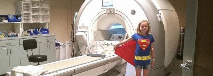
We suggest a baseline magnetic resonance imaging (MRI) or magnetic resonance angiography (MRA) test with and without contrast as soon as reasonable after birth or diagnosis, especially if the retinal blood vessels are abnormal by observations.
IP can be associated with learning disabilities. If your school-age child has delays or hardships despite a high IQ, have a neuro psychological exam. This will help you and educators at your school implement an individualized education program (IEP) so your child receives appropriate assistance.
Seizures or other complications should be treated as in any other infants.
Pulmonary
Recommended Specialist – Pulmonologist
IP can be associated with pulmonary hypertension. Have a non-evasive baseline test at birth and monitor closely for the first year of life. The suggested timeline is birth, three months, six months, nine months, and one year.
Genetics
Recommended Specialist – Geneticist
A geneticist should be and often is a family’s IP team leader for care throughout life, including follow-ups at the developmental milestones: pre-natal, all IP births, and puberty.
Molecular genetic (DNA) testing with or without skin biopsy is required for specific diagnosis of IP. Visit the CDC website for additional information on genetic testing and links to genetics experts.
Skin and Hair
Recommended Specialist – Dermatologist
A baby affected by IP should be watched carefully by a pediatric dermatologist for the first year of life.
Keep the skin cool and dry during all stages for the first year. It is imperative to avoid secondary infection. If it occurs, treat the secondary infection from common skin bacteria.
All stages of the skin may overlap one another for the first year of life and stages may repeat themselves. Each infant is different in severity and longevity. There is no “cure” for the skin stages. They will run their course for each case.
Associated with the scarring of the skin in IP, some people have had problems with heat intolerance and lack of sweating. If this is a concern, discuss this with your dermatologist. You can also download our Cooling Guide.
Teeth
Recommended Specialists – Pediatric Dentist, Dentist, Orthodontist, Oral Surgeon, Periodontist, Oral and Maxillofacial Surgeon
Recommended Dental Visits
Dental health is an important part of incontinentia pigmenti treatment. Routine dental care should be started by approximately 6 months of age to help prevent the likelihood of developing tooth decay and to begin evaluating the presence/absence of teeth. Dental development is monitored and preventive measures applied through early childhood. If missing and or malformed teeth are an esthetic issue for the child, treatment can be considered to help the child maintain a positive self-image.
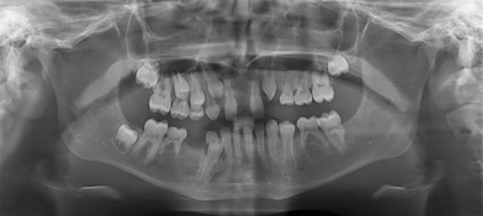
Missing and malformed teeth can be challenging to manage depending on several factors, such as, the number of teeth missing, the shape and location of the teeth present and the presence of tooth mal-alignment to mention just a few. Treatment may consist of bonding and crowns to reshape teeth, fixed or removable dentures or bridges to replacing missing teeth and implants. Orthodontics is commonly used to align teeth. A team approach is often best to manage these different oral issues and to help coordinate and time treatments optimally.